Imaging and Diagnostic Testing

Nuclear Imaging- Stress Test
Cardiac Nuclear Medicine uses a radioactive tracer called “Cardiolite” to produce an image of the heart and shows how well blood flows to the heart muscle. It is done in conjunction with an exercise stress test on a treadmill or can also be completed using medicines, such as Adenosine or Lexiscan, to simulate the effects of exercise on the heart. Cardiac Nuclear Medicine helps determine whether coronary artery stenosis (blockages) are so severe as to limit blood flow to the heart muscle when it needs it most, during physical activity. Cardiac Nuclear Medicine also determines the heart’s pumping function (ejection fraction).
At Alaska Heart & Vascular Institute, resting pictures are first obtained using a “tracer” agent. Next, stress is performed using treadmill exercise or medications. When a patient reaches his or her maximum level of exercise, or after simulating exercise with medications, a small amount of tracer is injected into a vein. The patient then lies down on a table under a camera that detects the energy emitted from the radioactive tracer and generates pictures (or scans) that reflect the heart’s blood flow both at rest and following stress. If a portion of the heart muscle is under-perfused (doesn’t receive a normal blood supply), a deficiency of tracer activity in that area will appear on the finished images as a “defect.” Some patients require this test to be done over two days.
Cardiac PET CT
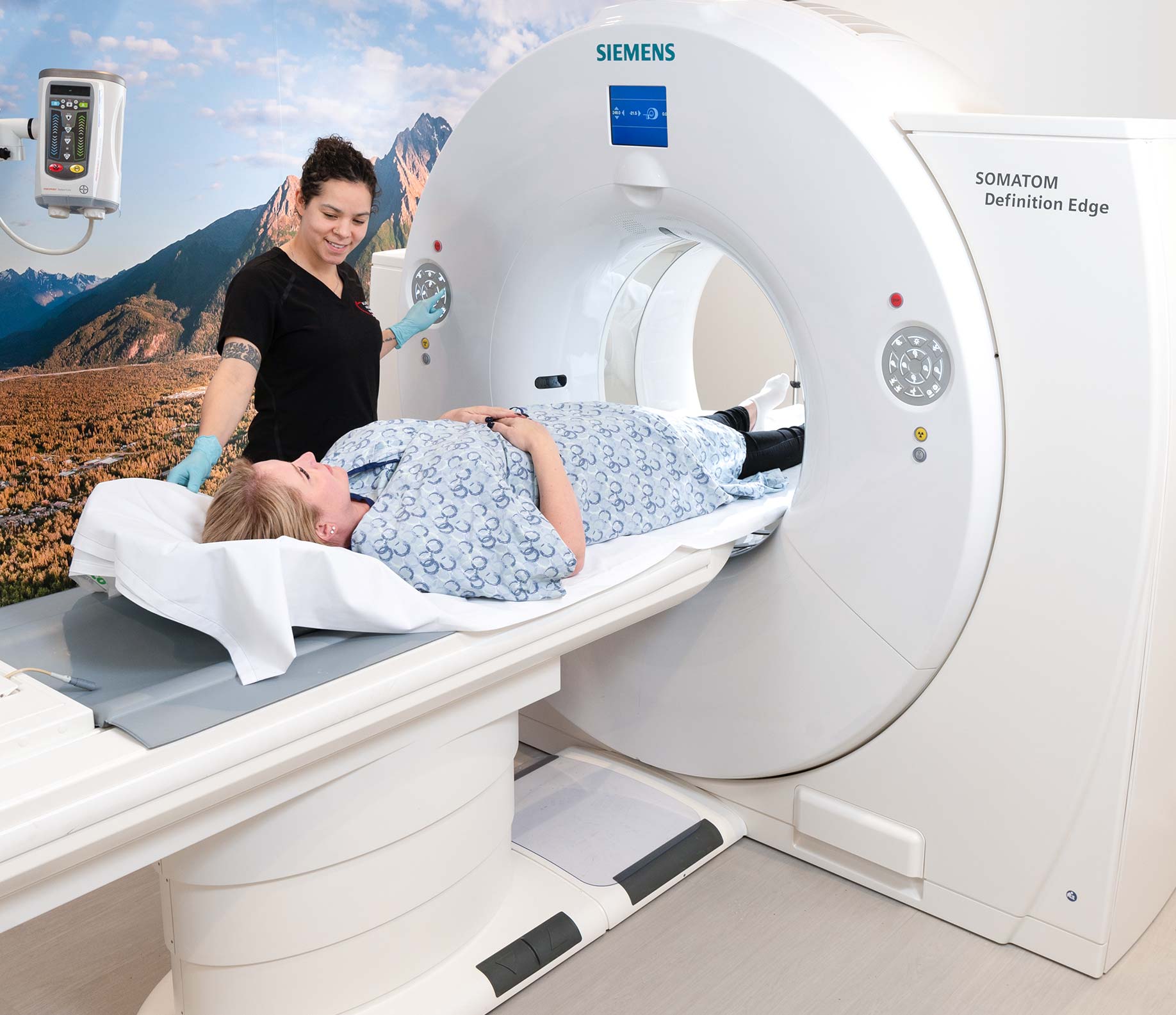
A baseline scan of your chest is obtained, followed by the “stress medication,” which takes the place of exercise. The second set of images is obtained, and the reading cardiologist can determine your heart function from the comparison of images. This entire test is performed on the PET CT table with no waiting between images.
Our “tracers” and methods are both widely used and safe. These tests take from 1-3 hours, and you may resume normal activity and diet immediately after. Cardiologists, nurses, exercise physiologists, and technologists with expertise in nuclear cardiology supervise and analyze these test results.
Computed tomography (CT) combines the use of X-rays with the latest computer technology. Using a series of X-ray beams, the CT scanner creates cross-sectional images. A computer reconstructs the “slices” to produce the actual pictures. Considering that some slices are as thin as half a millimeter, a CT scan offers more image detail than a traditional X-ray, which means your doctor gets the best information to make the most accurate diagnosis.
Magnetic resonance imaging (MRI) uses radiofrequency waves and a strong magnetic field rather than X-rays to provide remarkably clear and detailed pictures of internal organs and tissues. The procedure is valuable in diagnosing a broad range of conditions in all parts of the body, including heart and vascular disease, stroke, cancer, and joint and musculoskeletal disorders. MRI is unique in that it can also create detailed images of blood vessels without the use of contrast material, although there is a trend toward the use of special non-iodinated MRI contrast material; for example, gadolinium. MRI requires specialized equipment and expertise, and allows for the evaluation of particular body structures that may not be as visible with other imaging methods.
MRI is becoming very important in the initial diagnosis and subsequent management of coronary heart disease. MRI can help physicians to look closely at the structures and function of the heart and major vessels quickly and thoroughly, without the risks associated with traditional, more invasive procedures. Using MRI, physicians can examine the size and thickness of the chambers of the heart, and determine the extent of damage caused by a heart attack or progressive heart disease.
